Abstract
-
Background
Intramural esophageal dissection (IED) is a rare esophageal injury that is usually managed conservatively. However, the optimal treatment for complicated IED associated with infection or mediastinal abscess remains unclear.
-
Case
A 57-year-old man with decompensated liver cirrhosis presented with chest pain, odynophagia, and fever. Chest computed tomography revealed IED complicated by mediastinal abscess. Despite conservative management and thoracoscopic drainage, persistent fever suggested inadequate drainage of the infected false lumen. Endoscopic internal drainage was performed by longitudinal mucosal incision, followed by enteral nutritional support via feeding jejunostomy. The patient recovered without major surgery. Six-month follow-up endoscopy demonstrated complete healing of the false lumen with restoration of a single esophageal lumen.
-
Conclusions
Endoscopic internal drainage may be a feasible organ-preserving treatment option for selected patients with infected IED when adequate drainage cannot be achieved by conservative management or surgical drainage alone.
-
Keywords: Intramural esophageal dissection; False lumen; Mediastinal abscess; Endoscopic internal drainage; Liver cirrhosis; Case report
Introduction
Intramural esophageal dissection (IED) is a rare esophageal injury characterized by separation of the mucosal and submucosal layers, resulting in the formation of a false lumen [
1,
2]. Although most cases are managed conservatively and have a favorable prognosis, complicated cases associated with infection or mediastinal abscess are extremely uncommon, and their optimal management remains unclear [
2-
4]. We report a case of infected IED complicated by mediastinal abscess that was successfully treated with endoscopic internal drainage following surgical drainage while avoiding esophagectomy.
Case Report
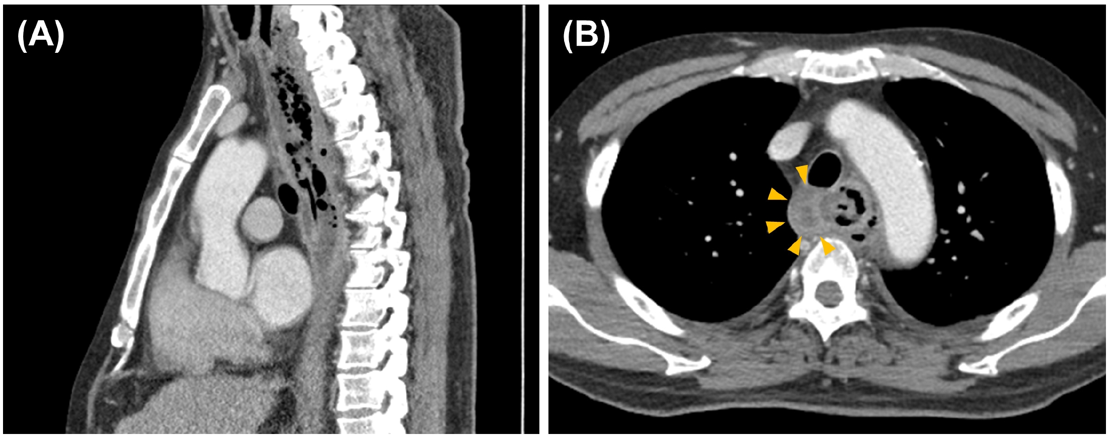
A 57-year-old man with alcohol-related liver cirrhosis complicated by esophageal varices, paroxysmal atrial fibrillation receiving direct oral anticoagulants, diabetes, and hypertension presented to the emergency department with worsening pain extending from the neck to the retrosternal area, odynophagia, and fever. On admission, his blood pressure was 127/75 mmHg, heart rate 81 beats/min, respiratory rate 20 breaths/min, and temperature 38.2°C. Laboratory findings showed a white blood cell count of 5,300/μL (74.0% neutrophils), hemoglobin of 12.9 g/dL, platelet count of 62×10³/μL, CRP of 87.5 mg/L, total bilirubin of 1.7 mg/dL, albumin of 3.1 g/dL, AST/ALT of 30/18 IU/L, and PT-INR of 1.51. Contrast-enhanced chest computed tomography (CT) revealed a linear intramural defect in the mid-thoracic esophagus with diffuse submucosal fluid collection, suggestive of intramural esophageal dissection (IED), accompanied by superior mediastinal abscess formation with extraluminal air, suggesting possible microperforation (
Fig. 1 A,
B). Small bilateral pleural effusions were also present.
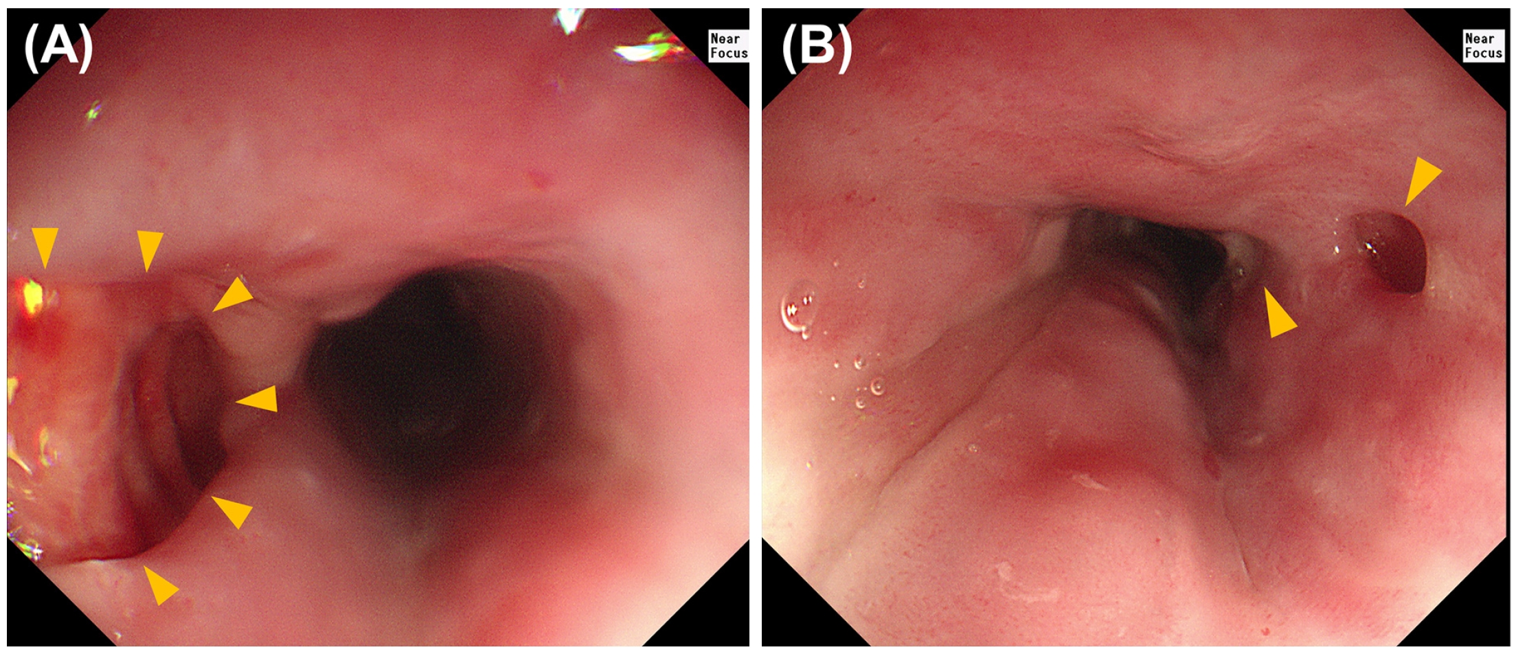
The patient was managed conservatively with bowel rest and intravenous ceftriaxone plus metronidazole. However, fever and chest pain persisted despite treatment. On hospital day 5, video-assisted thoracoscopic surgery was performed. A 1-cm abscess pocket above the azygos vein was identified and drained, and a right chest tube was inserted. No gross esophageal perforation or fistulous communication was identified. Upper endoscopy performed on hospital day 6 demonstrated diffuse edematous changes with multiple mucosal openings communicating with the intramural false lumen, from which blood-tinged purulent fluid was observed draining (
Fig. 2 A,
B). Grade I esophageal varices were also present.
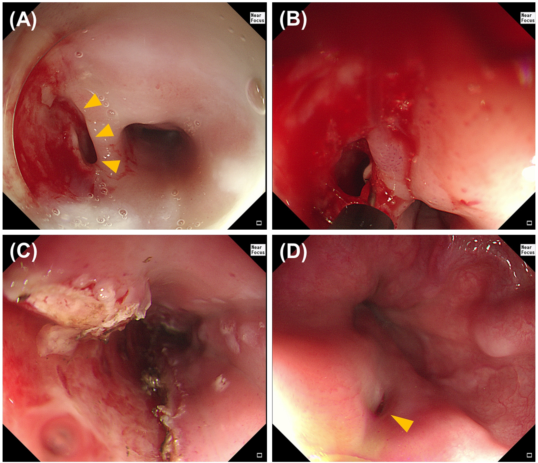
Although inflammatory markers gradually decreased after surgery, persistent fever raised concern for inadequate drainage of the infected intramural cavity. Therefore, therapeutic endoscopic intervention was planned on hospital day 10. Under general anesthesia, the largest mucosal opening was longitudinally extended from 28 to 35 cm from the upper incisors using an Insulated Tip knife to facilitate internal drainage (
Fig. 3 A-
D). Only minor transient oozing occurred during the procedure, without significant bleeding. Esophageal varices remained stable throughout the procedure. Further incision was not attempted because of marked mural edema, luminal narrowing, underlying coagulopathy, the presence of esophageal varices, and evidence of ongoing spontaneous drainage of purulent fluid through the distal mucosal openings. Because prolonged fasting was anticipated, a radiologically guided feeding jejunostomy was placed on hospital day 12, and enteral nutrition was initiated. After 17 days of intravenous antibiotics, the inflammatory markers markedly improved. Following chest tube removal, the patient was discharged with amoxicillin/clavulanate administered via the feeding jejunostomy.
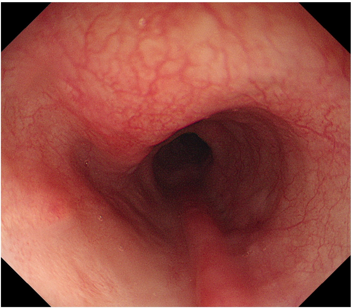
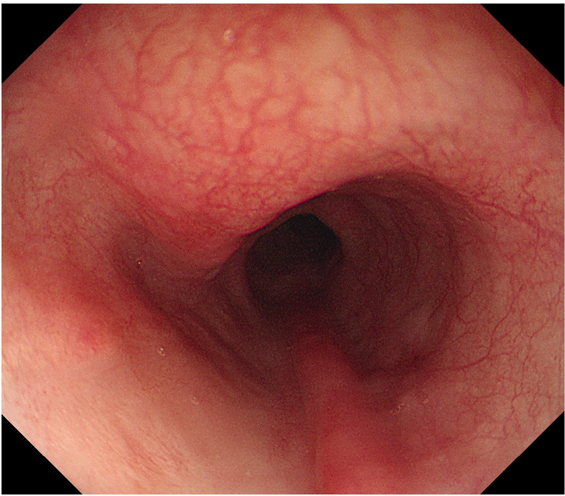
Serial chest CT examinations performed at 1- to 2-month intervals continued to suggest residual communication between the true and false lumens in the upper thoracic esophagus. During follow-up, the patient prematurely resumed oral intake against medical advice, resulting in transient mild aggravation of the radiologic findings. Therefore, conservative management with enteral nutrition and antibiotics was continued. After normalization of inflammatory markers and sustained clinical improvement, follow-up endoscopy was performed 6 months after the initial presentation. Endoscopy demonstrated complete epithelialization of the previous false lumen with restoration of a single patent esophageal lumen (
Fig. 4). Mild residual narrowing remained but did not impede endoscope passage. Oral feeding was resumed gradually without recurrence of symptoms.
Discussion
IED is a rare esophageal injury characterized by separation of the mucosal and submucosal layers, resulting in the formation of a false lumen [
1,
2]. It has been associated with esophageal instrumentation, forceful vomiting, coagulopathy, anticoagulant therapy, and underlying esophageal disorders [
1,
5]. Patients typically present with acute chest pain, odynophagia, or dysphagia, which may mimic more common cardiopulmonary conditions [
1,
2]. Contrast-enhanced CT and upper endoscopy play complementary roles in establishing the diagnosis by demonstrating the false lumen and excluding transmural perforation [
2,
4].
Most reported cases of IED follow a benign clinical course and respond well to conservative management consisting of fasting, nutritional support, acid suppression, and antibiotics when infection is suspected [
1,
3]. In contrast, complicated IED accompanied by secondary infection or mediastinal abscess is exceedingly uncommon, and the optimal management strategy has not been established [
4,
5]. Although surgical intervention may be required in patients with uncontrolled sepsis or suspected perforation, esophagectomy is a highly invasive procedure associated with substantial morbidity and mortality, particularly in patients with advanced liver cirrhosis or other significant comorbidities [
6,
7]. Therefore, organ-preserving treatment strategies are desirable whenever clinically feasible.
Our patient presented with infected IED, as evidenced by persistent fever, elevated inflammatory markers, purulent drainage from the false lumen, and concomitant mediastinal abscess formation. Despite thoracoscopic drainage of the mediastinal abscess, persistent fever suggested inadequate drainage of the infected false lumen. Endoscopic longitudinal mucosal incision successfully converted the narrow mucosal opening into a wider internal drainage tract, facilitating continuous drainage of the infected cavity [
8,
9]. Clinical and radiologic resolution was subsequently achieved without additional major esophageal surgery, suggesting that endoscopic internal drainage may serve as an effective adjunctive treatment when surgical drainage alone is insufficient [
8-
10].
In this case, decompensated liver cirrhosis complicated by esophageal varices, recurrent ascites, and hepatic encephalopathy was considered to confer a substantial perioperative risk for esophagectomy [
6,
7]. In this context, an organ-preserving approach was particularly desirable. To minimize the risk of bleeding, the mucosal incision was intentionally limited to the proximal lesion. Further distal extension was avoided because spontaneous drainage through multiple distal mucosal openings was already evident, while marked mural edema, luminal narrowing, and the presence of esophageal varices increased the anticipated procedural risk. The procedure was completed without clinically significant bleeding, and follow-up endoscopy demonstrated complete healing of the false lumen with restoration of a single esophageal lumen.
In conclusion, endoscopic internal drainage may represent a feasible organ-preserving treatment option for selected patients with infected IED, particularly when extensive surgical resection carries substantial operative risk [
6-
10]. Further studies are warranted to better define its role in the management of complicated IED.
Notes
Conflict of Interest
The authors declare no conflict of interest.
Funding
No funding was received for this work.
Data Availability Statement
Not applicable.
Ethics Approval and Consent to Participate
Institutional Review Board exemption was granted by the Institutional Review Board of Chung-Ang University Hospital (IRB No. 2608-001-19639), because written informed consent for publication could not be obtained due to loss to follow-up.
Authors' Contributions
Conceptualization: JYP. Patient management and investigation: JYP and BJP. Writing – original draft: JYP. Writing – review & editing: JYP, BJP, BJK, and JGK. All authors read and approved the final manuscript.
Fig. 1.Chest CT demonstrating (A) intramural esophageal dissection with (B) mediastinal abscess formation (arrowheads).
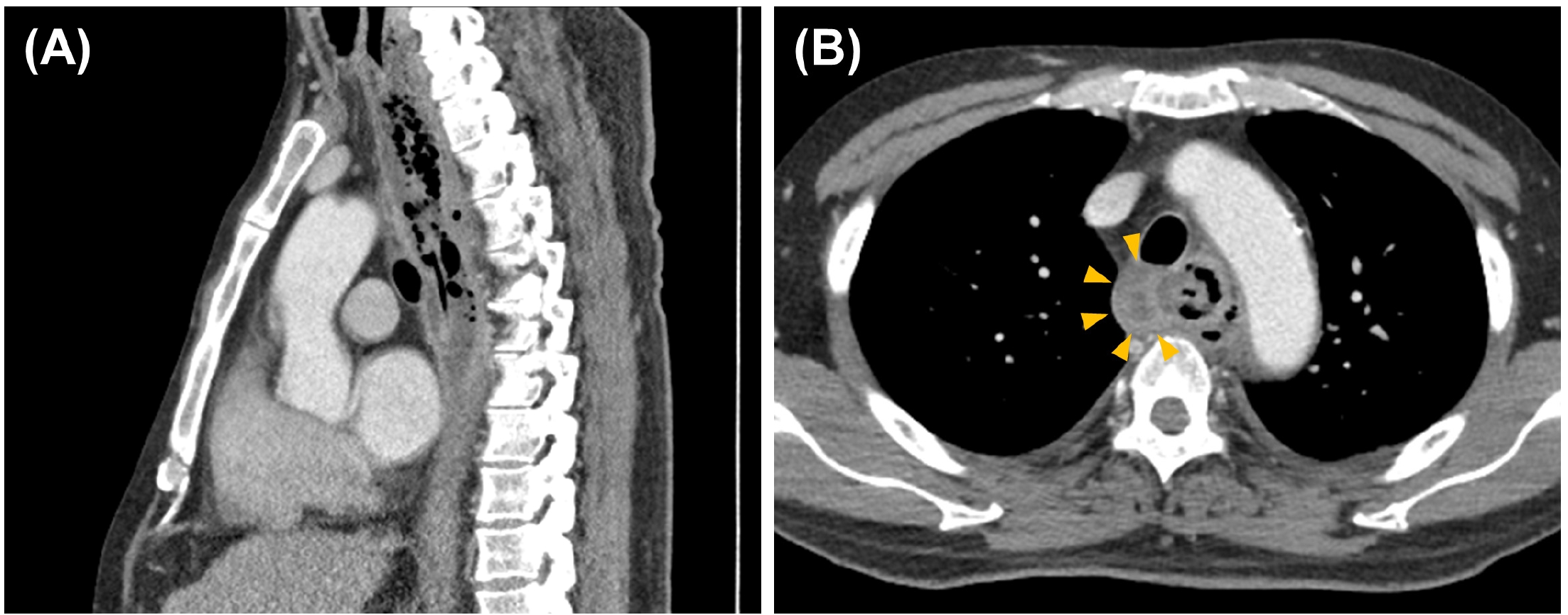
Fig. 2.Initial endoscopic findings. Upper endoscopy performed on hospital day 6 demonstrated diffuse edematous changes throughout the esophagus. (A) A large proximal mucosal opening (arrowheads; approximately 1.5 cm in diameter) located 28 cm from the upper incisors, communicating with a longitudinal intramural cavity (false lumen). (B) Multiple distal spontaneous mucosal openings with blood-tinged purulent drainage (arrowheads).
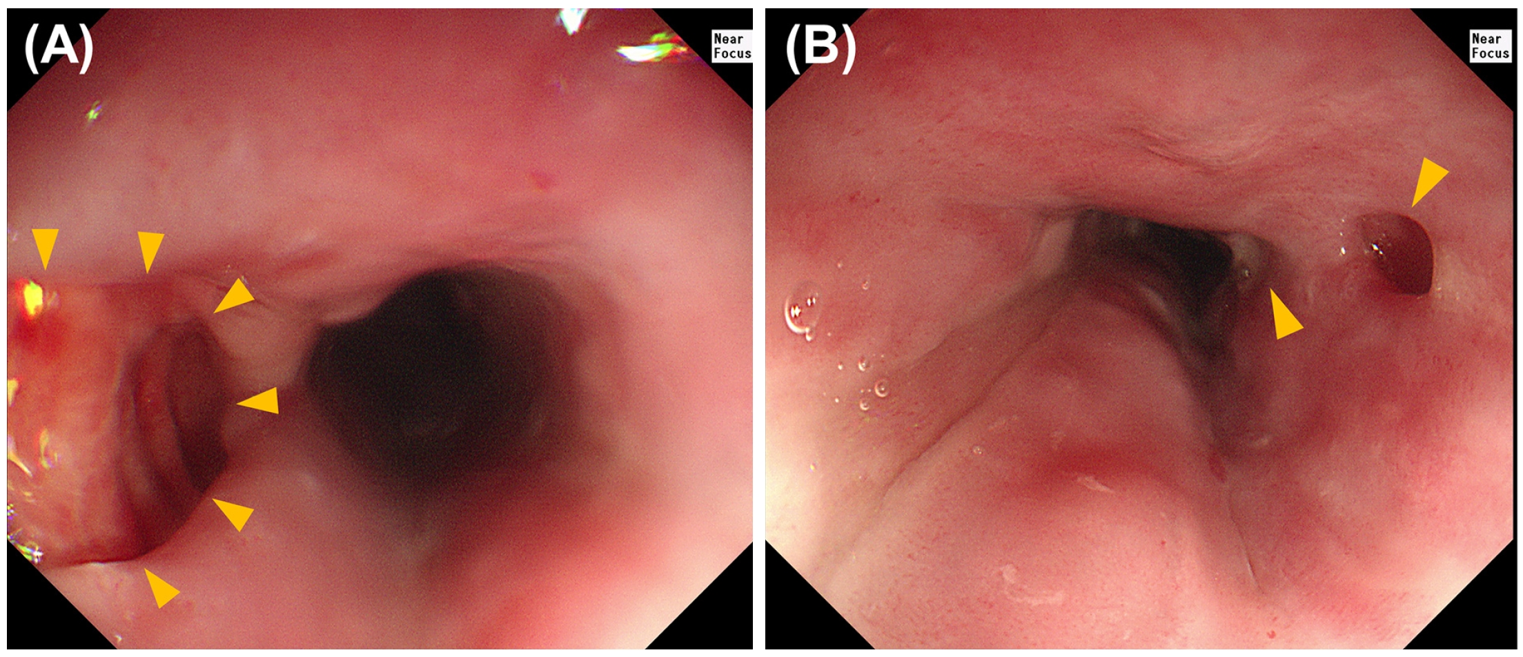
Fig. 3.Endoscopic internal drainage by longitudinal mucosal incision. (A) A large proximal mucosal opening at 28–29 cm from the upper incisors (9 o'clock position; arrowheads). (B) Longitudinal extension of the mucosal opening to approximately 35 cm using an Insulated Tip Knife to facilitate internal drainage. (C) Post-procedural appearance showing the widely opened false lumen with (D) persistent spontaneous drainage through multiple distal mucosal openings (arrowhead). No significant procedure-related bleeding was observed.
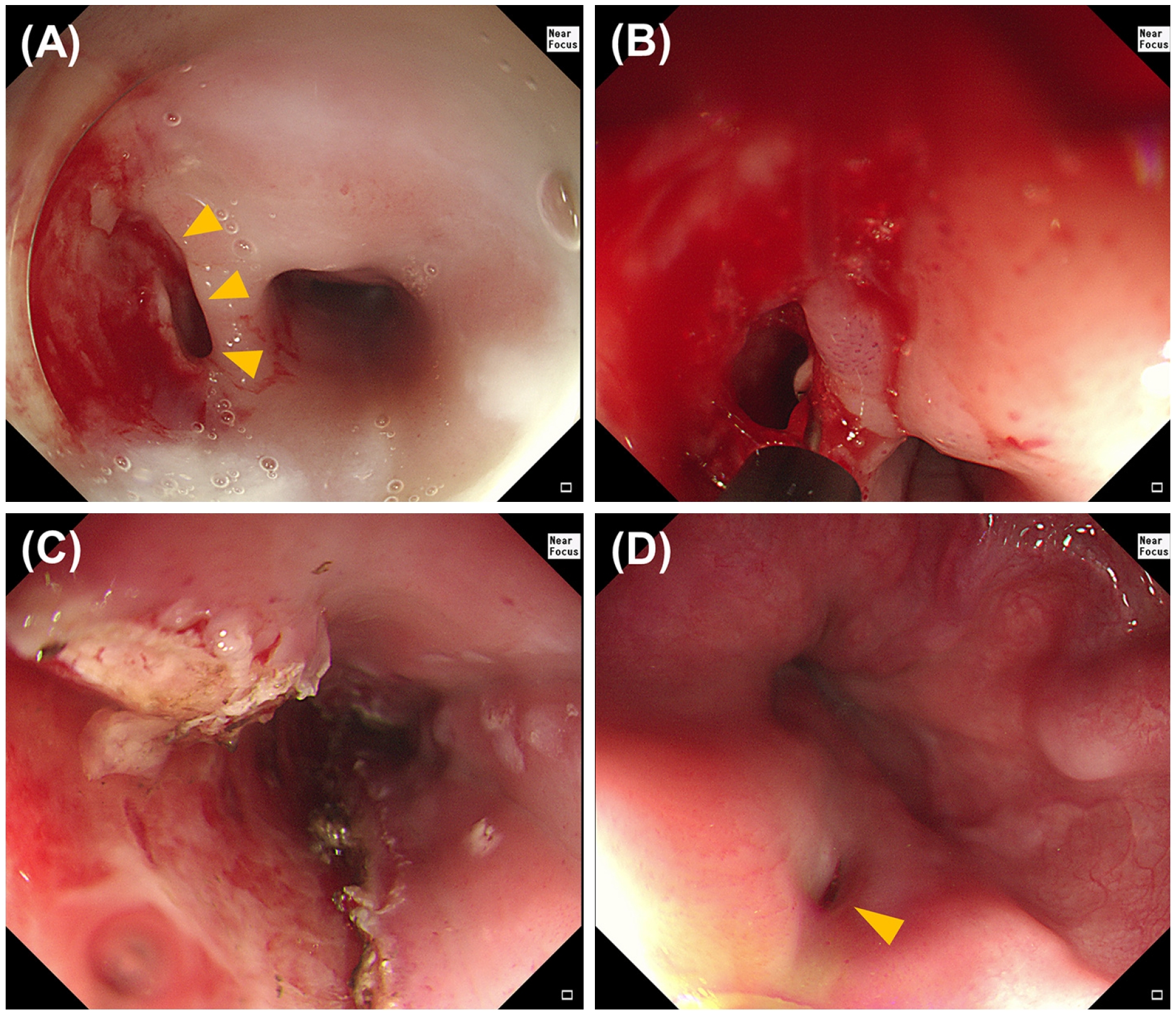
Fig. 4.Follow-up endoscopy 6 months after treatment showing complete epithelialization of the false lumen with restoration of a single esophageal lumen.
References
- 1. Beumer JD, Devitt PG, Thompson SK. Intramural oesophageal dissection. ANZ J Surg 2010; 80: 91-5.
- 2. Phan GQ, Heitmiller RF. Intramural esophageal dissection. Ann Thorac Surg 1997; 63: 1785-6.
- 3. Cheng G, Wang L, You J, Cheng L, Yang F, Wang X, et al. Asymptomatic intramural esophageal dissection: A case report and literature review. Medicine (Baltimore) 2025; 104: e42632.
- 4. Kumar S, Sakthivel MK, Bosemani T. Intramural Esophageal Abscess Complicated with Pleural Fistula: A Case Report. Cureus 2020; 12: e6846.
- 5. Bak YT, Kwon OS, Yeon JE, Kim JS, Byun KS, Kim JH, et al. Endoscopic treatment in a case with extensive spontaneous intramural dissection of the oesophagus. Eur J Gastroenterol Hepatol 1998; 10: 969-72.
- 6. Low DE, Kuppusamy MK, Alderson D, Cecconello I, Chang AC, Darling G, et al. Benchmarking Complications Associated with Esophagectomy. Ann Surg 2019; 269: 291-8.
- 7. Sozzi M, Siboni S, Asti E, Bonitta G, Bonavina L. Short-Term Outcomes of Minimally Invasive Esophagectomy for Carcinoma In Patients with Liver Cirrhosis. J Laparoendosc Adv Surg Tech A 2017; 27: 592-6.
- 8. Cho CM, Ha SS, Tak WY, Kweon YO, Kim SK, Choi YH, et al. Endoscopic incision of a septum in a case of spontaneous intramural dissection of the esophagus. J Clin Gastroenterol 2002; 35: 387-90.
- 9. Zhang J, Wang H, Liu D. Treatment of a spontaneous intramural esophageal abscess by endoscopic mucosal incision: an easy and effective therapy. Endoscopy 2022; 54: e937-8.
- 10. Benatta MA, Grimaud JC, Kaci M, Desjeux A, Baghdadi M, Loumi A. Intramural esophageal dissection due to pharyngeal abscess treated by endoscopic esophageal transection: a case report. Gastroenterol Clin Biol 2010; 34: 329-31.
Citations
Citations to this article as recorded by